
banner image

Conditions

From common eye concerns to complex conditions, Mr Gorman's specialist care is designed to preserve and improve your eyesight.

Cataracts

Cataract surgery is a safe, advanced procedure that replaces the eye’s clouded lens with a clear artificial lens to restore sharp, bright vision. 

A cataract is the clouding of the lens inside the eye.  The lens is situated behind the Iris (the coloured part of the eye).  The healthy lens is clear allowing light to the retina at the back of the eye.  The retina then transmits electrical signals to the optic nerve and into the brain.  The brain interprets this information into what we see. 
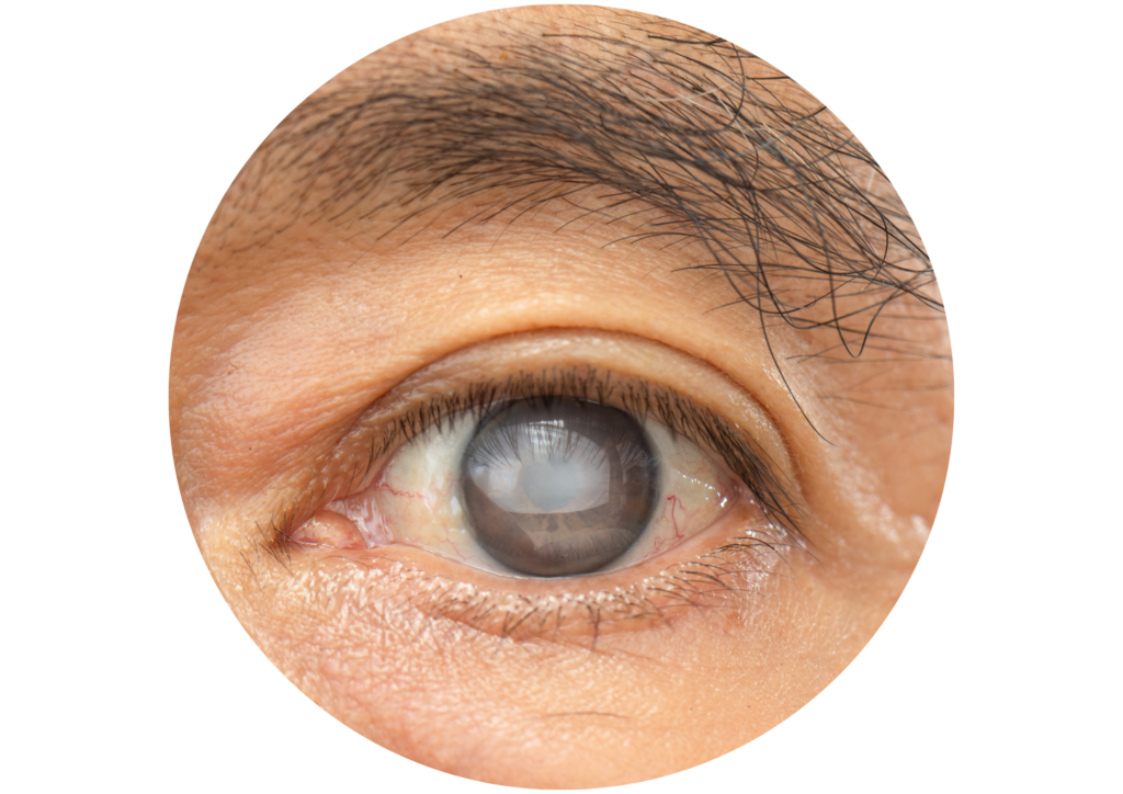
Cataracts develop due to changes in the substance of the lens and its water content.  As a result, over time the lens becomes cloudy.  Cataracts gradually progress causing symptoms such as: reduced vision, glare or light halos in certain lighting conditions, misty or clouded vision, blurring, double vision, colours not as vibrant and reduced light into the eye.   As the cataracts develop the reduced vision can affect the ability to carry out daily activities such as reading text and driving.  In severe cases cataract can cause blindness.

As cataracts are normally as a result of ageing, they are more common in older adults.  There are other factors that may increase the risk of developing cataracts including: previous eye injury or trauma, previous eye surgery, previous injections into the eye, other eye conditions including glaucoma and diabetes and long-term use of medications such as steroids.

A healthy natural lens has the capacity to change shape, enabling focus on objects near or far, which is known as ‘accommodation’.  With age, the lens accommodation changes and many people then need reading glasses to see close items.  This may be referred to as Presbyopia and is not cataract.

Posterior Capsular Opacification (PCO)

MANAGEMENT: YAG LASER CAPSULOTOMY

Following previous successful cataract surgery or clear lens extraction, patients may later on experience ‘clouding’ or blurry vision and potentially symptoms of increased visual glare or halo.  These symptoms can occur any time after cataract surgery in c. 20-25% of this patient population.  

During cataract surgery the natural lens of the eye is removed and replaced by a new artificial intraocular lens, however, the capsule (the bag-like structure that holds the lens) remains.  Cells can migrate and settle over the back (posterior) surface of the lens capsule causing a membrane thickening which prevents light effectively passing through to the retina at the back of the eye.  This is a natural attempt by the body’s healing system to repair the eye after surgery.  For the vast majority of patients, once the scar tissue has been removed with the YAG laser, light can pass again directly to the retina the vision improves.  PCO is not a return of the original cataract. 

Lorem ipsum dolor ist amte, consectetuer adipiscing eilt. Aenean commodo ligula egget dolor. Aenean massa. Cum sociis natoque penatibus et magnis dis parturient montes, nascetur ridiculus mus. Donec quak felis, ultricies nec, pellentesque eu, pretium quid, sem.

Retinal Detachment

Retinal detachment is when the retina separates from the supporting vessels and tissues underneath often due to the formation of a hole or tear which allows fluid to pass underneath the retina.  Retinal detachment is a sight threatening condition and usually requires urgent surgery

Symptoms include flashes of light, a shadow or curtain across the vision, large floaters, blurred vision and loss of vision.   Retinal detachment is normally painless. 

There are various risk factors that make retinal detachment more likely to occur including: severe myopia (short sightedness), eye trauma, previous eye surgery, natural ageing of the eye and family history of retinal detachment. 

The exact surgery depends on features of  the individual case.

Lorem ipsum dolor ist amte, consectetuer adipiscing eilt. Aenean commodo ligula egget dolor. Aenean massa. Cum sociis natoque penatibus et magnis dis parturient montes, nascetur ridiculus mus. Donec quak felis, ultricies nec, pellentesque eu, pretium quid, sem.

Macular Hole

The Macula is relatively small area of the central retina with the highest density, of the most light sensitive photoreceptors.  The centre of the macula is the fovea, which gives you your most precise sharp, detailed vision.  It is with this area that you read, watch TV and see detail.  You need it to be functioning well to drive a car. 

A macular hole is a small, circular gap or defect that develops in the macula, which unfortunately always centres on the fovea.  Central vision is badly affected by a macula hole.  Given time patients with an untreated macula hole will become unable to see even the top letter of the optician vision chart even with glasses.

Because the macula is responsible for the sharp, detailed central vision needed for tasks like reading, driving, and recognising faces, a hole in this area directly disrupts your straight-ahead line of sight.

Causes – most macula holes occur as an unfortunate side effect of aging of the vitreous in patients.  They usually present over the age of 60 and are more common in women. It is not usually found to be the result of a specific cause, although they can be associated with trauma and prior surgery.

  1. When should I see a retina specialist for a macular hole?

If you experience any blurring or distortion of central vision, or the presence of a shadow in your field of vision, then you need to seek immediate medical attention. Key Symptoms of a macula hole

  1. Distorted vision: Straight lines (like door frames or text) appear wavy, bowed, or crooked.
  2. Blurred central vision: Fine details become difficult to see clearly.
  3. Blind spots: As the hole expands, a small dark patch or “missing patch” appears right in the middle of your visual field.
  4. Preserved side vision: Peripheral (side) vision remains entirely unaffected, meaning it does not cause total blindness.
  5. Can a macular hole cause blindness?

Although macular holes do not usually lead to loss of peripheral vision, they have a great impact on central vision and thus make activities such as reading and driving challenging.

  1. How is a macular hole diagnosed?

A macular hole is usually first detected by an optician often after the patient becomes aware of poor vision on one eye.  The diagnosis is suspected just by looking at the back of the eye with special lenses during a thorough retinal examination.   A specialist imaging technique called an Optical Coherence Tomography (OCT) is often used to conform the diagnosis.

  1. Can a macular hole heal on its own?

Some very early-stage macular holes can close on their own, but the large majority cases of macular hole closure need surgery to restore vison and prevent further vision impairment.

  1. Is surgery usually successful?

Most holes picked up early can be closed with an operation.  Approximately 70% of patients who have surgery notice a very worthwhile improvement in vision.  Unfortunately, not all holes that close can be guaranteed to regain good vision, which  is therefore the biggest risk of surgery.  The sooner the operation is performed after you are aware of a problem, the more likely it will lead to a successful outcome.

Other complications of the surgery often include cataracts.  More serious complications are fortunately rare after macula hole surgery, but as with all retinal operations this still needs to be considered in your decision.

Lorem ipsum dolor ist amte, consectetuer adipiscing eilt. Aenean commodo ligula egget dolor. Aenean massa. Cum sociis natoque penatibus et magnis dis parturient montes, nascetur ridiculus mus. Donec quak felis, ultricies nec, pellentesque eu, pretium quid, sem.

Epiretinal Membrane (ERM)

Epiretinal membrane is a  thin layer of scar tissue forms across the macula.  This membrane can contract causing the retinal tissue underneath to distort. This may be referred to as cellophane maculopathy or macular pucker. Epiretinal Membranes are normally painless.  Symptoms may include distorted, wavy or reduced central vision 

Epiretinal Membrane may occur as part of the natural ageing process as the vitreous humour shrinks and pulls.  It may also occur due to inflammation, trauma or conditions such as Diabetic eye disease. 

The management of an epiretinal membrane (ERM) depends entirely on the severity of your symptoms and how much the condition interferes with your daily life. Because eye drops, medications, and updated glasses prescriptions cannot dissolve or correct the membrane, management ranges from routine observation for mild cases to surgical removal if vision is significantly impaired. 

Lorem ipsum dolor ist amte, consectetuer adipiscing eilt. Aenean commodo ligula egget dolor. Aenean massa. Cum sociis natoque penatibus et magnis dis parturient montes, nascetur ridiculus mus. Donec quak felis, ultricies nec, pellentesque eu, pretium quid, sem.

Diabetic Eye Disease

Diabetes, either requiring insulin, or treated with drugs only, can both lead to serious sight threatening eye disease.  Prevention and early detection is the first line of management. Good control of the diabetes and the blood sugar levels significantly reduce the risks of vision being affected.  Eyes should also be screened regularly in adult diabetics for signs of developing retinal problems.  Early treatment with laser and possibly injections can reduce the development of serious sight loss.  

Unfortunately, patients still develop serious sight threatening disease that may require vitrectomy.  Being referred for Vitrectomy surgery in diabetics is a sign of serious problems.  It is sadly by that stage often the only option for restoring and preserving vision. Patients referred for surgery to Mr gorman for the complications of Diabetic eye disease, have previously usually already been under the care of an ophthalmologist.  Normally it is that ophthalmologist who has requested his Mr Gorman’s help dealing with difficult disease progression.  Surgery may be advised to remove blood to improve vision, and to allow ongoing medical treatment of the retina, or may be directed at removing serios scarring form the retinal surface and flattening areas of retina that have been pulled of by the contracting scars.  Each case if judged individually.  Surgery for complex diabetic eye disease is sadly often very physically and emotionally demanding of the patient to undergo.  Thankfully the majority of patients do well, particularly given the context and presenting visions.    It is not however unusual that multiple surgeries and clinic visits may be required before the outcome is secured.

Lorem ipsum dolor ist amte, consectetuer adipiscing eilt. Aenean commodo ligula egget dolor. Aenean massa. Cum sociis natoque penatibus et magnis dis parturient montes, nascetur ridiculus mus. Donec quak felis, ultricies nec, pellentesque eu, pretium quid, sem.

Retinal Tear

A retinal tear is a break in the light-sensitive layer at the back of the eye—usually caused when the eye’s internal gel (vitreous) pulls on the tissue. It requires urgent medical evaluation within 24 to 48 hours for evaluation as left untreated a high proportion of retinal tears progress to retinal detachment.  Timely treatment with laser  can therefore prevent and detachment and the need for more complex vitrectomy surgery intervention. Laser is done in the eye clinic

Common Symptoms suggestive of  a retinal tear

  1. Sudden flashes of light in your side vision
  2. A sudden burst of new floaters, spots, or cobwebs
  3. Blurry or hazy vision
  4. A dark shadow or curtain moving across your field of vision

Main Causes and Risk Factors

  1. Aging: The vitreous gel naturally shrinks and pulls away from the retina.
  2. Short sightedness: High myopia stretches and thins the retinal tissue.
  3. Eye Trauma: Physical injury or past eye surgeries.
  4. Family History: A genetic predisposition to retinal problems

Lorem ipsum dolor ist amte, consectetuer adipiscing eilt. Aenean commodo ligula egget dolor. Aenean massa. Cum sociis natoque penatibus et magnis dis parturient montes, nascetur ridiculus mus. Donec quak felis, ultricies nec, pellentesque eu, pretium quid, sem.

eye surgery cardiff

SPECIALIST EXPERIENCE, PERSONALISED CARE

  • 0+ years

    Of consultant-led ophthalmic experience across NHS and private practice, diagnosing and treating a wide range of eye conditions.

  • 0+

    patients seen each week

Trusted by patients

Don John

“Lorem ipsum dolor sit amet, consectetur adipisicing elit, sed do eiusmod tempor incididunt ut labore et dolore magna aliqua.”

Don John

“Lorem ipsum dolor sit amet, consectetur adipisicing elit, sed do eiusmod tempor incididunt ut labore et dolore magna aliqua.”

Don John

“Lorem ipsum dolor sit amet, consectetur adipisicing elit, sed do eiusmod tempor incididunt ut labore et dolore magna aliqua.”

Don John

“Lorem ipsum dolor sit amet, consectetur adipisicing elit, sed do eiusmod tempor incididunt ut labore et dolore magna aliqua.”

Don John

“Lorem ipsum dolor sit amet, consectetur adipisicing elit, sed do eiusmod tempor incididunt ut labore et dolore magna aliqua.”