
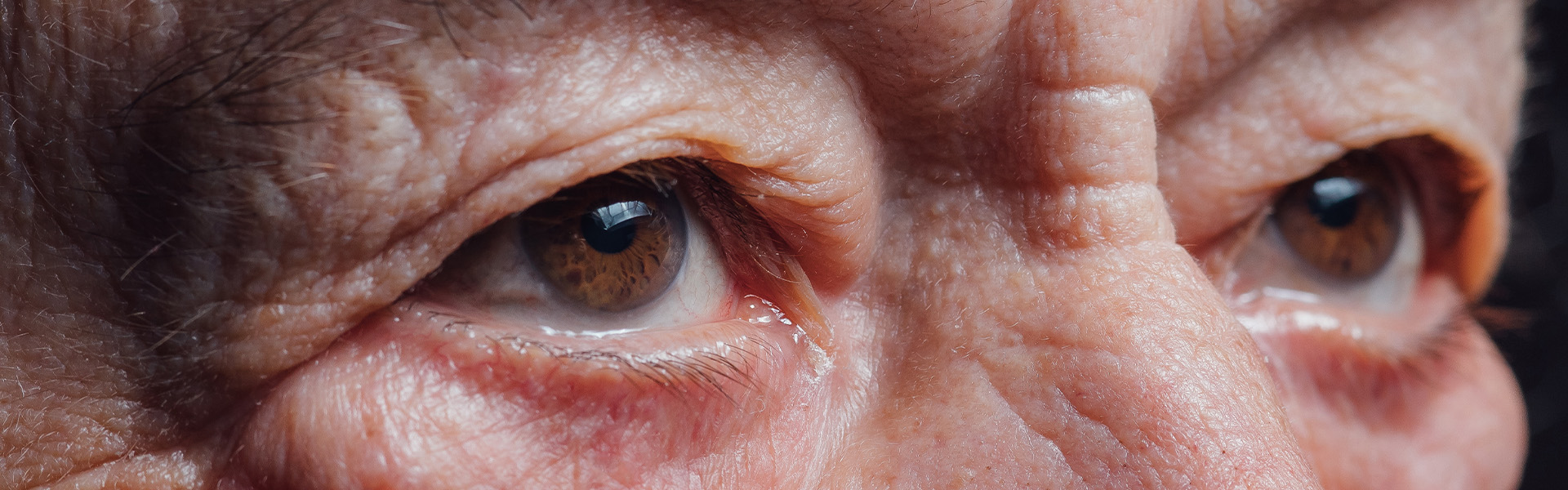




Cardiff Eyes

The treatments we offer

Mr Gorman is a highly experienced ophthalmic surgeon who manages a full range of general ophthalmic medical conditions. His specialist interests are in Cataract Surgery, Diabetic Eye Disease and the surgical treatment of Vitreoretinal disorders.

At Spire Cardiff Hospital, Mr Gorman personally manages each of his private patients.

Whatever the ophthalmic condition, a comprehensive eye examination and consultation is the starting point of a patient’s management.

Not every ophthalmic condition requires surgery but during a consultation at Spire Hospital, Mr Gorman has the time to explain his findings to the patient, discuss the condition, management options, timescales and answer patient questions.

Patients can then take time to consider their options and a management plan is agreed with the patient as appropriate.
eye exam
happy eyes

Common ophthalmic conditions that are seen in clinic but not necessarily requiring surgery include:

  • Allergy,
  • Blepharitis,
  • Diabetic Eye Disease,
  • Dry Eye,
  • Infection,
  • Inflammation.

Treatments

Cataract Surgery

  • The only treatment for a cataract is surgery. Surgery will normally be recommended when your visual symptoms begin to affect everyday activities such as reading or driving.

    During cataract surgery, the cloudy natural lens is removed using a technique called phacoemulsification. It is replaced with a clear artificial intraocular lens, known as an IOL. The artificial lens will normally remain in the eye for the rest of your life.

    The right time to have surgery differs from person to person. Mr Gorman will assess the general health of your eye and consider any other eye conditions that could affect the outcome. He will then explain the potential risks and benefits as they apply to you.

    What are the benefits of cataract surgery?

    For someone without another eye condition or additional risk factors, cataract surgery will usually produce a significant improvement in vision.

    Removing a cataract may also help with the management of other conditions:

    • It may help reduce pressure inside the eye in some patients with glaucoma.

    • It can make the retina easier to examine and monitor in patients with conditions such as age-related macular degeneration or diabetes.

    • It may reduce or eliminate the need for distance glasses.

    • It may help correct short-sightedness, long-sightedness or astigmatism.

    Cataract surgery cannot guarantee that every patient will be completely free from glasses. Mr Gorman will discuss the expected outcome during your consultation.

    Why choose private cataract surgery with Mr Gorman?

    • Continuity of care: Mr Gorman personally manages each stage of your care, from the initial consultation and surgery through to your postoperative review.

    • Direct contact: You can contact Mr Gorman’s private office if you have questions or concerns at any stage.

    • Convenience: Clinic appointments and surgery can usually be arranged without a lengthy wait.

    • Experience: Mr Gorman is an experienced cataract and vitreoretinal surgeon who works with an established team at Spire Cardiff Hospital.

    • Care for complex cases: His specialist experience allows treatment to be adapted for patients with more complex eye conditions or additional support needs.

    • Informed choice: You and, where appropriate, your family or carers will receive the information needed to make an informed decision. There is no pressure to proceed with surgery.

    • Postoperative care: Mr Gorman offers a face-to-face postoperative consultation following a patient’s first invasive surgical procedure.

    Assessment for cataract surgery

    An initial 30-minute consultation is required to determine whether cataract surgery is appropriate.

    Mr Gorman will examine both eyes, assess their general health and identify any conditions that could affect the result of surgery.

    Biometry will also be performed. This is a non-invasive test that measures the length and shape of the eye. These measurements are considered alongside your previous optical prescription, eye health and lifestyle requirements to help select the most appropriate lens.

    Please tell Mr Gorman if you have previously undergone laser vision correction, including LASIK or LASEK. These procedures alter the shape of the eye and must be considered when calculating the lens power.

    Before your assessment, stop wearing contact lenses for:

    • At least five days if you wear soft contact lenses.

    • At least two weeks if you wear hard or gas-permeable contact lenses.

    This allows the eye to return to its natural shape before measurements are taken.

    Additional investigations, such as corneal mapping or topography, may occasionally be required.

    Lens options

    Monofocal lenses

    Monofocal lenses are the most commonly used lenses in cataract surgery. They provide clear vision at one focal distance, which is often set for distance vision and driving.

    Reading glasses or glasses for close work will usually still be required.

    Multifocal lenses

    Multifocal lenses divide light between different focal zones. They are designed to provide vision at several distances, including near, intermediate and distance vision.

    These lenses are not suitable for everyone. They may be unsuitable for patients with conditions including:

    • Macular degeneration or macular scarring.

    • Previous retinal or vitrectomy surgery.

    • Amblyopia.

    • Corneal disease.

    • Significant dry eye.

    Possible compromises include:

    • Glare, starbursts or halos, particularly at night.

    • Reduced contrast in dim lighting.

    • A longer period of visual adaptation.

    • The continued need for glasses at certain distances.

    Multifocal lenses are custom ordered and normally cost more than monofocal lenses.

    Extended depth of focus lenses

    Extended depth of focus lenses stretch the range of focus rather than splitting light into separate zones. They may provide a smoother transition between different distances and better contrast in low light.

    Reading glasses may still be required. Mr Gorman will discuss these lenses if they are suitable for you.

    Toric lenses

    Toric lenses are designed for patients with astigmatism. Astigmatism occurs when the eye has an uneven curvature, causing light to focus at more than one point.

    A toric lens is shaped to compensate for this curvature. These lenses are usually custom ordered and generally cost more than monofocal lenses.

    Mr Gorman will discuss the lens option he recommends for your eye and lifestyle.

    What are the risks of cataract surgery?

    Cataract surgery is generally safe, but no operation is completely free from risk. Most complications are uncommon and can be treated or managed, although further treatment or surgery may sometimes be required.

    Possible risks include:

    • Bruising around the eye.

    • Vitreous loss.

    • Part of the natural lens moving into the back of the eye.

    • Bleeding around or inside the eye.

    • Damage to the cornea, iris or lens capsule.

    • Infection inside the eye, known as endophthalmitis.

    • Inflammation or swelling at the macula.

    • Retained lens material.

    • A temporary increase in pressure inside the eye.

    • Movement of the implanted lens.

    • An increase in noticeable floaters.

    • Corneal swelling.

    • Rejection of a previous corneal graft.

    • Retinal detachment.

    • Drooping of the eyelid, known as ptosis.

    • Reactivation of an existing condition such as diabetes, macular degeneration or herpes infection.

    • Glare, halos or a shadow effect caused by the artificial lens.

    • Blurred or reduced vision.

    • The continued need for glasses to refine the final result.

    Mr Gorman will explain how these risks apply to your individual eye before surgery.

    Can a cataract grow back?

    The artificial lens will not cloud in the same way as the natural lens, so the cataract itself cannot return.

    In approximately 20–25% of patients, the capsule behind the artificial lens may gradually become cloudy. This is known as posterior capsular opacification and can develop at any time after cataract surgery.

    It can usually be treated with a short outpatient laser procedure called a YAG capsulotomy.

    Additional support requirements

    Please tell Mr Gorman during your initial consultation if you think you may need additional assistance before, during or after surgery.

    This may include:

    • Fear of surgery or significant anxiety.

    • Hearing difficulties.

    • Claustrophobia.

    • Difficulty lying flat or remaining still.

    • Breathing difficulties.

    • Dementia or cognitive impairment.

    • Mobility difficulties or the need for a hoist.

    • A head tremor or another physical condition.

    Mr Gorman and the hospital team will assess your requirements and make appropriate arrangements wherever possible.

    Preparing for cataract surgery

    Cataract surgery is normally performed as a day-case procedure under local anaesthetic. You should expect to remain at the hospital for a morning or afternoon.

    Before surgery:

    • Arrange for someone to take you home, as you must not drive after the operation.

    • Stop wearing contact lenses for at least five days before surgery, unless advised otherwise.

    • Make sure high blood pressure is well controlled.

    • If you have diabetes, make sure your blood glucose is well controlled.

    • Keep your eyelids and eyelashes clean.

    • Do not wear eye makeup during the days immediately before surgery.

    • Make sure conditions such as blepharitis are treated before the operation.

    • Tell Mr Gorman about all allergies, including a latex allergy.

    • Confirm with the clinical team how to manage your usual medication.

    Most prescribed medicines can be continued normally. However, some medicines, including anticoagulants or blood thinners, may need to be stopped temporarily. Only change your medication when instructed by your clinical team.

    If a general anaesthetic is required, the hospital team will tell you when to stop eating and drinking.

    On the day of surgery

    • Attend the main Spire Cardiff Hospital building rather than Glamorgan House.

    • Wear comfortable clothing that does not need to be pulled over your head, such as a button-front shirt or cardigan.

    • If you are having local anaesthetic, you can normally drink as usual and have a light meal before attending hospital.

    • Drops will be used to dilate your pupil before surgery. This may take some time.

    • The operation itself normally takes approximately 20 minutes.

    • You will be asked to lie flat, and a sterile drape will be placed over the eye area.

    • After surgery, you will be given time to rest and will normally be offered refreshments.

    • You will be discharged once the hospital team is satisfied that it is safe for you to return home.

    A clear plastic eye shield will be placed over the operated eye. Do not remove it until the following morning unless the clinical team gives you different instructions.

    You will also receive postoperative eye drops and written care instructions.

    Caring for your eye after surgery

    The operation is performed through a small incision. While it heals, the eye needs to be protected from infection and injury.

    The incision usually heals within approximately 7–10 days. Most patients experience a straightforward recovery.

    Activities to avoid

    During the early recovery period:

    • Do not rub or touch the operated eye.

    • Avoid heavy lifting and strenuous exercise.

    • Do not swim or use a hot tub or sauna for one month.

    • Avoid environments with a higher risk of dirt or infection, such as gyms and gardening areas.

    • Take care not to get dust, dirt, soap or other products in the eye.

    • Wear glasses or other suitable protection in windy conditions.

    • Avoid applying makeup around the eye until your postoperative review.

    Activities you can continue

    You can normally:

    • Bend as usual.

    • Wash your face carefully, avoiding soap, dirty water and cleansing products around the operated eye.

    • Shower and wash your hair while protecting the eye from water and shampoo.

    • Watch television.

    • Use a computer.

    • Read.

    • Walk and remain mobile.

    • Eat normally and maintain good control of diabetes or high blood pressure.

    Always follow the personalised instructions provided by your clinical team.

    Eye shield

    Keep the eye shield in place until the morning after surgery unless instructed otherwise.

    When it is removed, the eye may initially feel sticky or crusted and the eyelids may be difficult to open. Gently clean around the eye using the wipes or saline supplied by the hospital. Boiled and cooled water may also be used with a clean, non-fibrous wipe if necessary.

    You will not normally need to wear the shield during the day after this. Wear it at night for the first week and, where possible, sleep on the non-operated side.

    Eye drops

    You will be given eye drops and instructions explaining when to use them. The drops help control inflammation and protect the eye against infection.

    • Use the drops exactly as prescribed.

    • Continue using them for the instructed period and until the prescribed course is complete.

    • If you already use prescribed eye drops for another condition, open a new bottle and continue using them unless advised otherwise.

    • Leave time between different eye drops so that one does not wash the other out.

    • Use a new, unopened bottle if you restart lubricating drops after surgery.

    Contact the hospital team or Mr Gorman’s office if you are unsure how to use your drops.

    Discomfort and pain relief

    Mild discomfort, itching or a gritty sensation can occur after surgery. These symptoms should improve rather than become worse.

    Paracetamol may be taken if appropriate for you. Significant or increasing pain is not expected and should be reported promptly.

    Restarting medication

    If you were instructed to stop medication before surgery, including an anticoagulant or blood thinner, follow the clinical team’s instructions about when to restart it.

    What to expect during recovery

    Temporary visual disturbance

    As the anaesthetic wears off, you may experience double, distorted or disturbed vision. This should settle as the muscles around the eye return to normal.

    Dilated pupil

    The pupil may remain dilated for up to 24 hours. This can cause blurred vision and sensitivity to light. Sunglasses may help.

    Redness

    Some redness in the white of the eye is common when the eye shield is first removed. It should gradually improve.

    Colours and brightness

    Colours may appear brighter or more vibrant after the cataract has been removed. This is because the cataract previously created a yellow or sepia tint and reduced the amount of light entering the eye.

    Driving

    Do not drive until:

    • The anaesthetic has worn off.

    • Your pupil has returned to its normal size.

    • There is no double vision.

    • Your visual field is normal.

    • You can meet the current DVLA eyesight requirements.

    Your optician can confirm whether your vision meets the legal standard for driving.

    Glasses and contact lenses

    Your glasses prescription will probably change after surgery. Wearing your previous glasses will not damage the eye, but they may no longer provide clear vision.

    Wait at least three weeks, or for the period recommended by Mr Gorman, before arranging a new prescription.

    Do not wear a contact lens in the operated eye until Mr Gorman confirms that it is safe.

    Follow-up care

    • Mr Gorman or Alison will normally telephone you from lunchtime onwards on the day after surgery to check that you are comfortable.

    • If there are any concerns, Mr Gorman will arrange an examination.

    • Arrange an appointment with your optician approximately three to four weeks after surgery for a postoperative prescription or refraction.

    • OCT scans and visual-field tests are not routinely required unless specifically requested.

    • Ask your optician for a written copy of your new prescription to bring to your postoperative consultation.

    • Mr Gorman will confirm your follow-up arrangements. A clinic appointment is normally arranged approximately four to five weeks after surgery.

    If you have questions or concerns, contact Alison on the private office telephone number: 029 2048 5847.

    When to seek urgent advice

    Contact Mr Gorman’s office promptly if you experience:

    • Pain: More than mild discomfort or pain that is becoming worse.

    • Worsening vision: A reduction in vision after an initial improvement.

    • New floaters: A sudden increase in the number of floaters.

    • A curtain-like shadow: A dark shadow moving across your vision.

    • Increasing redness: Redness that becomes more pronounced rather than settling.

    If you are concerned about any new or worsening symptom, contact Mr Gorman’s office on 029 2048 5847.

Vitreous and retinal surgery

A vitrectomy is an eye operation that removes the clear, gel-like substance from the centre of the eye, known as the vitreous gel. It can be a sight-saving procedure.

Removing the vitreous allows the surgeon to access and treat conditions affecting the retina at the back of the eye. The operation may also be used to remove blood, debris or scar tissue that is obstructing vision.

Sometimes blood within the vitreous prevents the retina from being examined properly. In these circumstances, a vitrectomy may be needed to identify and treat a potentially serious retinal condition before it causes permanent damage to vision.

Why might a vitrectomy be needed?

A vitrectomy may be recommended for conditions including:

  • Retinal detachment: To reposition and secure retinal tissue that has pulled away from the back of the eye.
  • Diabetic retinopathy: To remove blood from the vitreous or scar tissue caused by diabetes.
  • Macular hole: To help close a small hole in the macula, the central part of the retina responsible for detailed vision and reading.
  • Epiretinal membrane: To remove scar tissue that is wrinkling or distorting the macula.
  • Eye trauma: To remove foreign material or clear severe bleeding inside the eye.
  • Complications of cataract surgery: To stabilise the eye and treat problems involving the vitreous or retina.
Your consultation

Mr Gorman has more than 25 years’ experience as a consultant performing vitrectomy surgery and caring for patients with complex retinal conditions.

Before recommending surgery, he will examine your eye and discuss your symptoms, general health, other eye conditions and any factors that could affect the outcome.

The potential benefits and risks will be explained in relation to your individual circumstances, helping you make an informed decision about whether surgery is right for you.

What happens during the operation?

Most vitrectomy procedures are performed under local anaesthetic. An anaesthetist numbs the eye using an injection around it, and the eye usually remains numb for several hours.

During surgery, Mr Gorman creates three small entry points in the white part of the eye, known as the sclera. Using a microscope and specialist microsurgical instruments, he may:

  • Remove cloudy vitreous, blood or debris.
  • Remove scar tissue from the retina.
  • Remove foreign material from inside the eye.
  • Reposition the retina against the back of the eye.
  • Use laser treatment to repair a retinal tear or treat another retinal problem.
  • Place an air or gas bubble in the eye to support the retina while it heals.
  • Place silicone oil in the eye, which may need to be removed during a later operation.

Vitrectomy surgery usually takes approximately 45–90 minutes. It is normally performed as a day-case procedure, allowing you to return home after surgery unless other arrangements have been made.

After the operation

Before leaving hospital, you will receive instructions about caring for your eye. You will normally be prescribed eye drops, which may include:

  • Anti-inflammatory drops.
  • Antibiotic drops.
  • Drops to manage pressure inside the eye, if required.

Vision will usually be blurred for several weeks after surgery, particularly when a gas bubble has been placed inside the eye. Vision should gradually improve during the following weeks and months, although recovery varies according to the condition being treated.

Posturing after surgery

If a gas bubble is used, you may need to keep your head in a particular position after the operation. This is known as posturing.

The required position and length of time will depend on your condition and the procedure performed. Mr Gorman and his team will provide specific instructions.

Posturing helps the gas bubble act like a supportive splint against the treated area of the retina during the early healing period. You may be asked to maintain the required position for approximately 50 minutes of every hour, followed by a 10-minute break.

Posturing can be demanding, and you may need help at home during this period.

Important instructions when a gas bubble is used

While a gas bubble remains inside your eye:

  • Do not fly or travel to a high altitude until Mr Gorman confirms that it is safe. Reduced atmospheric pressure can cause the bubble to expand, creating a dangerous rise in pressure inside the eye.
  • Tell your anaesthetist about the gas bubble if you require an operation under general anaesthetic. Certain anaesthetic gases can cause the bubble to expand and increase pressure inside the eye.
  • Follow all posturing instructions carefully.
  • Use your prescribed eye drops as directed.
  • Contact the clinical team promptly if you have any concerns.
Follow-up care

Mr Gorman will normally review you in the clinic:

  • The day after surgery.
  • Approximately two weeks after surgery.
  • Approximately eight to ten weeks after surgery.

Additional appointments may be required depending on your recovery.

These examinations allow Mr Gorman to monitor the health of the eye, assess the progress of the operation and identify any potential complications. Your follow-up plan will be personalised to your needs, and you will be given contact details in case you have concerns.

Benefits of vitrectomy surgery

The aim of vitrectomy surgery is usually to improve vision, prevent further deterioration or preserve the health of the eye.

The likely benefit depends on the condition being treated, its severity and other factors affecting the eye. Some patients may require more than one operation, particularly if a retina detaches again or a macular hole does not close following the first procedure.

Mr Gorman will explain the expected benefits in your individual case before asking you to consent to surgery.

What are the risks?

Vitrectomy is a significant eye operation. Although it is generally safe, complications can occur and some may threaten vision.

Possible risks include:

  • Cataract: Vitrectomy can accelerate clouding of the eye’s natural lens, particularly in people over 50, and cataract surgery may later be required.
  • Retinal detachment: The retina may pull away from the back of the eye for the first time or detach again after previous treatment.
  • Infection: A rare but serious infection inside the eye, known as endophthalmitis, requires urgent treatment.
  • Bleeding: Bleeding may occur inside the eye during or after surgery.
  • High or low eye pressure: Changes in pressure can lead to conditions such as glaucoma or hypotony.
  • Gas or silicone oil effects: These can cause temporary blurred vision and may require specific positioning or travel restrictions.
  • Inflammation: Swelling, redness or discomfort may occur on the surface or inside the eye.
  • Reduced vision: Vision may not improve as expected and, rarely, may become worse.
  • Further surgery: Another operation may be necessary if the original problem persists or a complication develops.

Mr Gorman will explain how these risks and potential benefits apply to your eye so that you can make an informed decision about surgery.

Lorem ipsum dolor ist amte, consectetuer adipiscing eilt. Aenean commodo ligula egget dolor. Aenean massa. Cum sociis natoque penatibus et magnis dis parturient montes, nascetur ridiculus mus. Donec quak felis, ultricies nec, pellentesque eu, pretium quid, sem.

YAG Laser Capsulotomy

Posterior capsular opacification, known as PCO, is treated with a procedure called YAG laser capsulotomy or posterior capsulotomy.

YAG laser capsulotomy is a quick and commonly performed treatment. It is completed as an outpatient procedure in a clinic rather than an operating theatre.

The procedure is non-invasive and does not create a wound in the eye.

Appointment booking advice

If your optician has advised that you have PCO and require YAG laser treatment, telephone Alison, Mr Gorman’s Secretary, directly on 02920 485847 to arrange an appointment.

Please contact Alison rather than Spire Cardiff Hospital Appointments because the YAG laser is located separately from the main outpatient clinic.

Where possible, a single appointment will be arranged to include both your initial consultation and the YAG laser procedure. This helps reduce the number of hospital visits and avoids the need to have your pupils dilated on separate occasions.

Preparing for your appointment

  • No formal preparation is required.

  • Eat and drink normally before your appointment.

  • Your pupils will need to be dilated. Arrange appropriate transport and do not drive to or from the appointment.

  • Bring your current glasses.

  • Bring sunglasses, as your eyes may be sensitive to light until your pupils return to their normal size.

  • Bring a copy of your optician’s referral or most recent glasses prescription and give it to Mr Gorman.

  • The consultation and procedure will usually take approximately 45 minutes in total, whether one or both eyes require treatment.

What happens at the appointment?

Mr Gorman will first examine both eyes to assess their condition and identify any other eye problems.

If YAG laser treatment is appropriate, he will explain the procedure and discuss its potential benefits and risks with you. YAG laser capsulotomy is generally considered a low-risk ophthalmic procedure, although complications can occur.

Drops will be placed in your eye to dilate the pupil. Anaesthetic eye drops will then be used to numb the surface of the eye. These drops may sting briefly.

You will be asked to sit at the laser machine with your chin resting on the chin support to help keep your head still.

Mr Gorman will place a small contact lens against the surface of the eye and carefully direct the laser beam towards the cloudy capsule behind the artificial lens.

The laser creates a small opening in the cloudy membrane, allowing light to pass through to the retina again.

The procedure normally takes only a few minutes and is not usually painful.

Recovery after YAG laser treatment

After the procedure:

  • Your eye may feel slightly dry, gritty or uncomfortable.

  • Your vision may remain blurred while the pupil is dilated.

  • Your eye may be sensitive to light until the pupil returns to its normal size.

  • You may notice floaters that appear as dots, lines or cobweb-like shapes.

Floaters are small clumps within the vitreous gel that fills the eye. They are commonly noticed after the procedure and usually become less noticeable over time, although some may persist.

Many patients notice an improvement in vision shortly after treatment. Temporary discomfort, blurred vision and light sensitivity should normally improve within 24 hours.

Aftercare

Eye drops are not usually required following YAG laser capsulotomy, and there are generally no specific activity restrictions. You can normally return to your usual daily activities.

Follow any personalised instructions given to you by Mr Gorman.

Your glasses prescription may need to be updated before you resume driving. If you are uncertain whether your vision meets the required standard, arrange an assessment with your optician.

Follow-up care

A routine follow-up appointment with Mr Gorman will only be arranged when there is a clinical need.

If you experience unusual symptoms or have any questions or concerns following the procedure, contact Alison, Mr Gorman’s Secretary, on 02920 485847.

Lorem ipsum dolor ist amte, consectetuer adipiscing eilt. Aenean commodo ligula egget dolor. Aenean massa. Cum sociis natoque penatibus et magnis dis parturient montes, nascetur ridiculus mus. Donec quak felis, ultricies nec, pellentesque eu, pretium quid, sem.

Retinal Laser Treatment

Retinal tear

A retinal tear will usually require urgent treatment to reduce the risk of it progressing to a retinal detachment.

When the tear has not progressed too far, it may be treated using one of the following procedures:

  • Laser retinopexy or photocoagulation: A specialist laser creates small, controlled areas of treatment around the retinal tear. As these areas heal, scar tissue forms and helps secure the retina, reducing the risk of fluid passing through the tear.

  • Cryopexy or cryoretinopexy: A freezing technique is applied to the outside of the eye over the affected area. This also creates scar tissue that helps secure the retina around the tear.

Cryopexy may be used when laser treatment cannot be performed adequately or when the location of the retinal tear makes laser treatment difficult.

Diabetic retinopathy

Diabetic retinopathy may be classified as:

  • Non-proliferative diabetic retinopathy (NPDR).

  • Proliferative diabetic retinopathy (PDR).

In more complex stages of diabetic eye disease, a specialist laser treatment called panretinal photocoagulation (PRP) may be used.

PRP treats areas of the retina affected by abnormal or newly formed blood vessels. Its purpose is to stabilise the eye and reduce the risk of further severe sight loss.

Mr Gorman regularly performs this treatment as an outpatient procedure.

Because diabetic retinopathy can continue to progress, laser treatment may not always be sufficient. In some cases, vitrectomy surgery may subsequently be recommended.

Lorem ipsum dolor ist amte, consectetuer adipiscing eilt. Aenean commodo ligula egget dolor. Aenean massa. Cum sociis natoque penatibus et magnis dis parturient montes, nascetur ridiculus mus. Donec quak felis, ultricies nec, pellentesque eu, pretium quid, sem.

General Ophthalmology

General ophthalmology

In addition to his specialist surgical interests in cataract surgery, vitreoretinal conditions and posterior capsular opacification, Mr Gorman also sees patients who require an opinion on more general eye symptoms and guidance about their management.

Symptoms assessed by Mr Gorman

Mr Gorman regularly sees patients experiencing:

  • Flashes and floaters.

  • Eye discomfort or pain.

  • Sensitivity to light, known as photophobia.

  • Poor eyesight or reduced vision.

  • Redness of the eye.

Ongoing advice and management

If a possible diagnosis has already been made, Mr Gorman can also provide ongoing advice about:

  • Conjunctivitis.

  • Blepharitis.

  • Dry eye.

  • Eyelid margin disease.

Lorem ipsum dolor ist amte, consectetuer adipiscing eilt. Aenean commodo ligula egget dolor. Aenean massa. Cum sociis natoque penatibus et magnis dis parturient montes, nascetur ridiculus mus. Donec quak felis, ultricies nec, pellentesque eu, pretium quid, sem.

Trusted by patients

Don John

“Lorem ipsum dolor sit amet, consectetur adipisicing elit, sed do eiusmod tempor incididunt ut labore et dolore magna aliqua.”

Don John

“Lorem ipsum dolor sit amet, consectetur adipisicing elit, sed do eiusmod tempor incididunt ut labore et dolore magna aliqua.”

Don John

“Lorem ipsum dolor sit amet, consectetur adipisicing elit, sed do eiusmod tempor incididunt ut labore et dolore magna aliqua.”

Don John

“Lorem ipsum dolor sit amet, consectetur adipisicing elit, sed do eiusmod tempor incididunt ut labore et dolore magna aliqua.”

Don John

“Lorem ipsum dolor sit amet, consectetur adipisicing elit, sed do eiusmod tempor incididunt ut labore et dolore magna aliqua.”